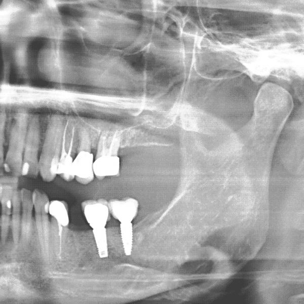
DIGITAL X-RAY
We have various diagnostic options available in the dental practice. If it is found during a clinical examination that the tooth defect is large and there is damage to the tooth root or tooth pulp, X-rays are taken. This also applies among other things to gingivitis, as the bone substance can also be affected.
Furthermore, digital images are taken in order to be able to clearly diagnose caries sites in the interdental spaces. These are not always visible to the naked eye.
X-ray diagnosis allows us to obtain information about the condition of the teeth, the roots of the teeth and the jawbone and is therefore essential in dentistry.
Patients are informed about the necessity of x-rays and shots are only taken if there is urgency.
As far as the radiation exposure is concerned, X-rays in a dental practice are classified as very low. The dose, measured in microsievert (μSv), depends on the mode of administration and the technique used. In our practice, we have a small-format X-ray device for dental film recordings and an ortho-tomography device including ceph arm to take an overview of your teeth and jaws. The radiation dose of these devices is between 0.2 and max. 8μSv. We X-ray with low radiation with digital or imaging plate technology.
Just for comparison and for your understanding, during a flight to the Canary Islands you will be charged with about 20μSv of cosmic radiation. For more information, visit http://www.bfs.de.
Digital X-ray technology dispenses with chemicals such as developer fluids and fixers.
Digital X-ray images are available after only a short time and can be shown to the patient clearly and understandably. In addition, it is possible to move the images internally to the respective treatment units and to forward them externally to colleagues.


